Digestive System Structured Answer Biology Class-9 ICSE Selina Publishers Solutions Chapter-11. Step By Step ICSE Selina Concise Solutions of Chapter-11 Digestive System with Exercise-11 including MCQs, Very Short Answer Type, Short Answer Type, Long Answer Type and Structured/Application Questions Solved . Visit official Website CISCE for detail information about ICSE Board Class-9.
Digestive System Exe-11 Structured Answer Biology Class-9 ICSE Concise Selina Publishers
| Board | ICSE |
| Publications | Selina Publication |
| Subject | Biology |
| Class | 9th |
| Chapter-11 | Digestive System |
| Book Name | Concise |
| Topics | Solution of A. Structured/Application/Skill Answer Type |
| Academic Session | 2023-2024 |
A. Structured/Application/Skill Answer Type
Digestive System Class-9 Biology Concise Solutions
Page 121
Question 1.
Draw a labelled diagram to show the internal structure of a mammalian tooth with two roots.
Answer:
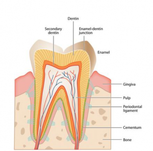
Internal structure of a mammlian tooth
Question 2.
Complete the following table by filling in the blanks 1 to 8.
| Organ | Enzyme | Food
acted upon |
Find
product |
| 1 | Pepsin | 2 | 3 |
| Mouth | 4 | 5 | Di
saccharide |
| 6 | 7 | Maltose | 8 |
Answer:
| Organ | Enzyme | Food
acted upon |
Find
product |
| Stomach | Pepsin | Proteins | Poly
peptide |
| Mouth | Amylase | Starch | Di
saccharide |
| Ileum | Maltase | Maltose | Glucose |
Question 3.
Study the diagram given below and then answer the questions that follow:
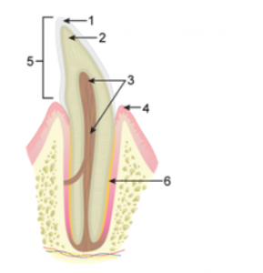
(a) Name the parts labelled 1, 2, 3, 4, 5 and 6.
(b) Identify the tooth and give a reason to support your answer.
(c) Describe the structure of the part labelled ‘3’.
(d) Give the total number of the type of tooth mentioned in ‘1’ above, in the mouth of an adult and state its function.
Answer:
(a)
1 → Enamel
2 → Dentine
3 → Pulp
4 → Gum
5 → Crown
6 → Cement
(b)
The tooth shown in the diagram has only one root, so it is an incisor or canine which is used for biting and piercing.
(c)
The part labelled ‘3’ (pulp) is a soft connective tissue present in the pulp cavity of the tooth. It consists of blood capillaries, lymph vessels and nerve fibres. These extend from the crown of the tooth and open through the pulp cavity at the base of the root.
(d)
Type of teeth in the mouth of an adult:
- Incisors (8) → Used for biting and cutting
- Canines (4) → Used for holding and tearing of food
- Premolars (8) → Used for grinding and crushing of food
- Molars (12)→ Used for grinding and crushing of food
Question 4.
Study the following dental formula and then answer the questions that follow:
i 3/4 c 0/0 pm o/1 m 1/1
(a) What is the total number of teeth in the (i) upper jaw and (ii) lower jaw?
(b) State the total number of teeth present in the dentition.
(c) Give the dental formula of an adult human being.
Answer:
(a) The total number of teeth in the upper jaw are 8 and lower jaw are 12.
(b) A total of 20 teeth are present in the given dentition.
(c) Dental formula of an adult human being is.
2,1,2,3
2,1,2,3
A. Structured/Application/Skill Answer Type
Digestive System Class-9 Biology Concise Solutions
Page 122
Question 5.
The figures (A, B, C and D) shown below represent different kinds of teeth in humans. Study the figures and answer the following questions:
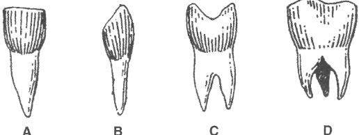
(a) What kind of teeth do A, B, C and D represent ?
(b) Write one structural feature/shape of each.
(c) Mention the number of teeth of each kind in one jaw with their specific position.
(d) Name two minerals present in teeth.
(e) What do you mean by ‘Wisdom tooth’?
Answer:
(a)
- A → Incisor
- B → Canine
- C → Premolar
- D → Molar
(b)
- A → Broad and sharp
- B → Conical and sharply pointed
- C → Two hill-like projections(bicuspid)
- D → Large grinding surface
(c)
- A → Four, centre of each jaw
- B → Two, one on each side of incisor
- C → Four, two on side of each canine
- D → Six, three on each side
(d) Calcium and phosphorous.
(e) Last molar on each side of jaw is called wisdom teath.
Question 6.
Draw a neat diagram of the “Microscopic Structure of an intestinal villus” and label the parts given below :
(i) Epithelium
(ii) Capillaries
(iii) Lacteals
Answer the following questions :
(a) What is the advantage of having a large number of villi on the inner surface of small intestine ?
(b) Write the important role of lacteals.
(c) Name the juice secreted from the glandular cells of small intestine.
(d) Name the specific secretions of:
(1) Salivary gland
(2) Stomach
(3) Liver
(4) Pancreas
Answer:
Below is the labelled diagram of the “Microscopic Structure of an intestinal villus”:
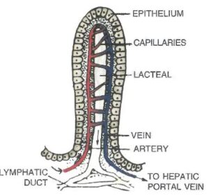
(a) A large number of villi enormously increase the inner surface area of the small intestine which facilitates the absorption of digested food.
(b) The fatty acids and glycerol are absorbed into the lacteals to enter the lymphatic system which forms a network all over the body to ultimately empty its contents into the blood stream.
(c) Intestinal juice
(d)
(1) Salivary gland – Saliva (amylase)
(2) Stomach – Gastric juice
(3) Liver – Bile
(4) Pancreas – Pancreatic juice
— : End of Digestive System E. Structured/Skill Answer Class-9 ICSE Biology Solutions :–
Return to Return to Concise Selina ICSE Biology Class-9
Thanks
Please share with your friends