The Nervous System and Sense Organs Class-10 Structured Questions Goyal Brothers ICSE Biology Solutions Ch-10. We Provide Solutions of Structured Questions of Exercise- 10 The Nervous System and Sense Organs. All solutions are given as council prescribe guideline for next upcoming exam. Visit official Website CISCE for detail information about ICSE Board Class-10 Biology.
Ch-10 The Nervous System and Sense Organs Structured Questions
| Board | ICSE |
| Publications | Goyal Brothers publications |
| Subject | Biology |
| Class | 10th |
| Writer | Dr. K.K. Aggrawal |
| Chapter-10 | The Nervous System and Sense Organs |
| Topics | Solutions of Structured Questions |
| Edition | for 2022-2023 Academic Session |
E. STRUCTURED QUESTIONS
Ch-10 The Nervous System and Sense Organs Goyal Brothers Prakashan ICSE Class-10 Biology Solutions
(Page-152)
Question 1. Given below is the diagram of the human ear. Study the same and answer the questions that follow :
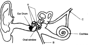
(i) Give the biological term for the part labelled ‘A’ and state its function.
(ii) Name the part labelled ‘B’ and state its function.
(iii) Name the part labelled ‘C’ and state its function.
(iv) Give the function of ear wax.
Answer :
(i) Part labelled ‘A’ is called as ear ossicles. It is made up of three bones malleus, incus and stapes. These ear ossicles transmit the sound waves from external to the internal ear.
(ii) Part labelled ‘B’ is called as eustachian tube. It acts as ventilator to equalize pressure of air on both the sides of tympanic membrane that forms the outer boundary of middle ear.
(iii) Part labelled ‘C’ is auditory nerve (vestibular and cochlear nerve) which carries hearing impulses to the brain.
(iv) Ear wax lubricates the tympanum for proper functioning.
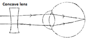
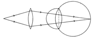
Question 2. Given below is a diagram depicting a defect of the human eye, study the same and then answer the questions that follow :
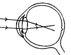
(i) Name the defect shown in the diagram.
(ii) What are the two possible reasons that cause this defect ?
(iii) Name the type of lens used to correct this defect.
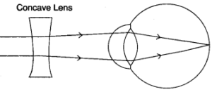
(iv) With the help of a diagram show how the defect shown above is rectified using a suitable lens.
Answer :
(i) Myopia.
(ii) 1. The eye ball is too long from front to back.
2. The lens is too curved.
(iii) Concave lens
(iv)
Question 3. The diagram given below is a representation of a certain phenomenon pertaining to the nervous system. Study the diagram and answer the following questions :
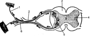
(i) Name the phenomenon that is being depicted.
(ii) Give the technical term for the point of contact between the two nerve cells.
(iii) Name the parts 1, 2, 3 and 4.
(iv) Write the functions of parts 5 and 6.
(v) How does the arrangement of neurons in the spinal cord differ from that of the brain ?
Answer :
(i) Reflex action.
(ii) Synapse
(iii) 1. Sensory/Afferent neuron.
2. Dorsal ganglion/Dorsal root.
3. White matter
4. Grey matter
(iv) Function of 5 (Synape): Transmit the sensory impulse from sensory neuron to the motor neuron.
Function of 6 (Motor neuron/Efferent neuron): Transmit the command to the effectors (muscle or glands).
(v) In Brain: Gray matter on the outerside and white matter on inner side.
In Spinal cord: White matter on the outerside and Gray matter on inner side.
Question 4. The diagram shows a section of the human brain. Answer the questions that follow :
(i) Name the parts labelled A, B and C.
(ii) Give the main function of each of the parts A, B, and C.
(iii) Name the three protective membranes covering the brain.
(iv) Name the basic unit of the brain.
Answer :
(i) Part A = Cerebrum
Part B = Cerebellum
Part C = Medulla oblongata.
(ii) Cerebrum: It is the site of controlling memory, reasoning, thinking, perception, emotions and speech.
Cerebellum: It maintains posture equilibrium and muscular co-ordination.
Medula Oblongata: It contains centre for cardiac, respiratory and vasomotor activities. It also co-ordinates reflexes for swallowing, coughing, sneezing and vomiting.
(iii) Three protective membranes covering the brain are : duramater, piamater and arachnoid.
(iv) The basic unit of brain is neuron.
Question 5. The diagram given along side represents the structure found in the inner ear. Study the same and then answer the questions that follow :
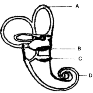
(i) Name the parts labelled A, B C and D.
(ii) Name the part of the ear responsible for transmitting impulses to the brain.
(iii) Name the part labelled above which is responsible for:
1. Static equilibrium.
2. Dynamic equilibrium.
3. Hearing.
(iv) Name the audio receptor cells which pick up vibrations.
(v) Name the fluid present in the inner ear.
Answer :
(i) Part A = Semicircular canals.
Part B = Utriculus
Part C = Sacculus
Part D = Cochlea
(ii) Auditory Nerve.
(iii) 1. Utriculus and sac culus
2. Semicircular canals.
3. Cochlea.
(iv) Organ of corti.
(v) Endolymph.
Question 6. Given below is a diagram depicting a defect of the human eye. Study the same and then answer the questions that follow :
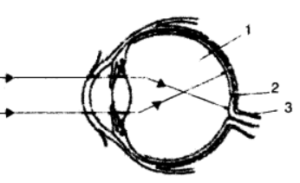
(i) Identify the defect.
(ii) Name the parts labelled 1, 2, and 3.
(iii) Give two possible reasons for this eye defect.
(iv) Draw a labelled diagram to show how the above mentioned defect is rectified.
Answer :
(i) Myopia.
(ii) Part labelled 1 – Vitreous Humor
Part labelled 2 – Yellow spot
Part labelled 3 – Optic Nerve
(iii) Two reasons for this defect are as follows :
1. The eyeball becomes long from front to back.
2. The lens becomes too curved convex.
(iv) This defect is to be rectified by Concave lens
Question 7. Study the following diagram carefully and then answer the questions that follow. The diagram is depicting a defect of the human eye :
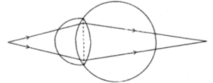
(i) Identify the defect shown in the diagram.
(ii) Give two possible reasons for the above defect.
(iii) Draw a neat labelled diagram to show how the above defect can be rectified.
Answer :
(i) Hypermetropia
(ii) (a) The eye ball is too short from front to back.
(b) The lens is too flat.
(iii)
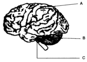
Question 8. The diagram given below is an external view of the human brain. Study the same and answer the questions that follow :
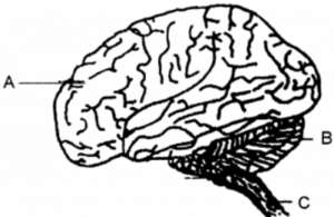
(i) Name the parts labelled A, B and C in the diagram.
(iii) What are the structural and functional units of the brain? How are the parts of these units arranged in A and C?
(iv) Mention the collective term for the membranes covering the brain.
(v) What is the function of Cerebrospinal fluid?
Answer :
(i) A. Cerebrum
B. Cerebellum
C. Spinal cord
(ii) A. It is the seat of memory, will power, emotions, experience, intelligence and controls all voluntary actions of the body.
B. Maintains balance of the body.
(iii) Neuron/Nerve cell.
In A grey matter is outside and white matter is inside while in C grey matter is inside and white matter is outside.
(iv) Meninges.
(v) It serves as shock absorbing medium. Protects brain and the rest of CNS against jerk and jolts. It maintains constant pressure in and around the brain.
— : End of The Nervous System and Sense Organs Structured Questions Class-10 Goyal Brothers Prakashan ICSE Biology Solutions :–
Return to :- ICSE Biology for Class 10 Goyal Brothers Prakashan solutions
Thanks