Sense Organs Class 10 Structured / Applications Concise Biology Selina Solutions Ch-11. In this article you will get the solutions / Answer of Structured / Applications / Skill Questions / Problems as council latest prescribe guideline. Visit official website CISCE for detail information about ICSE Board Class-10 Biology.
Sense Organs Class 10 Structured / Applications Concise Biology Selina Solutions Ch-11
| Board | ICSE |
| Subject | Biology |
| Class | 10 |
| Book | Selina Concise |
| Chapter-11 | Sense Organs |
| Topics | Solutions of Structured / Applications / Skill Type Questions |
Solutions of Structured / Applications / Skill Type Questions
Page 150-151
Que-1: The figures (A) and (B) given below are showing some kind of adjustment. Study the figures and answer the questions that follow.
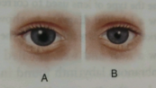
(a) Identify the kinds of adjustments done in the figure (A) and (B).
(b) Distinguish between the adjustments of figures (A) and (B) on the basis of :
(i) The size of pupil.
(ii) The pigment which gets regenerated.
(iii) Cells of the retina.
Ans:
(a) Kinds of adjustments done in the figure:
(A) dilated pupil due to dim light.
(B) Constricted pupil due to bright light.
(b)
| S. No. |
Factor | A | B |
|---|---|---|---|
| (i) | The size of pupil | bigger | smaller |
| (ii) | The pigment which gets regenerated | Rhodopsin (visual purple) | Iodopsin. |
| (iii) | Cells of the retina | Rods become active and cones become inactive | Cones become active and rods become inactive |
Que-2: With reference to human eye and ear answer the questions that follow :
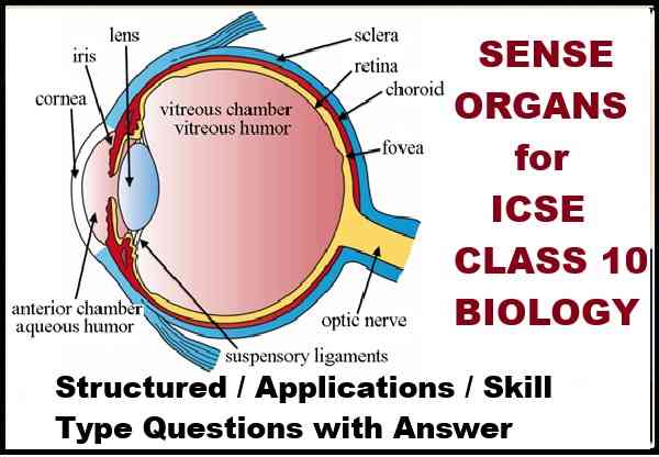
(a) Name the parts of the eye associated with:
(i) Regulation of the size of pupil.
(ii) Regulation of the shape of lens.
(iii) Keeping the lens moist and protecting it from physical shock.
(iv) The layer providing nourishment to the eye.
(b) Name the part of the ear associated with :
(i) Static balance.
(ii) Dynamic balance.
(iii) Hearing.
(iv) Amplification of vibrations.
Ans:
(a)
(i) Regulation of the size of pupil: Iris.
(ii) Regulation of the shape of lens: Ciliary muscles.
(iii) Keeping the lens moist and protecting it from physical shock: Aqueous Humour.
(iv) The layer providing nourishment to the eye: Choroid layer.
(b)
(i) Static balance: Vestibule.
(ii) Dynamic balance: Ampulla.
(iii) Hearing: Organ of Corti.
(iv) Amplification of vibrations: Ear ossicles (Malleus, incus and stapes).
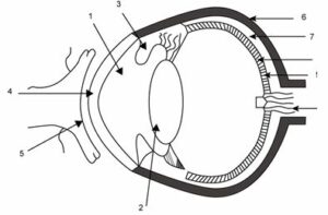
Que-3: The figure given below refers to the vertical section of the eye of a mammal. Study the figure carefully and answer the following questions.
(a) Label the guidelines shown as 1 to 10.
(b) Write one important role of parts shown as 3 and 7.
(c) Write one structural difference between the parts shown as 9 and 10.
(d) Mention one functional difference between the parts shown as 6 and 8.
Ans:
(a) The guidelines are labelled below:
1: Aqueous chamber
2: Lens
3: Iris
4: Cornea
5: Conjunctiva
6: Sclera
7: Choroid
8: Retina
9: Yellow spot
10: Optic nerve (Blind spot)
(b) Part 3 (Iris) — It contains radial muscles to dilate the pupil and circular muscles to constrict the pupil.
Part 7 (Choroid) — It is the middle layer of the eyeball, richly supplied with blood vessels and provides nourishment to the eye.
(c) Part 9 (yellow spot) contains sensory cells especially the cone cells while part 10 (blind sport) contains no sensory cells.
(d) Part 6 (sclera) gives shape to the eyeball and part 8 (retina) acts as screen to form image of an object.
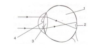
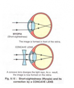
Que-4: Given below is a diagram depicting a defect of the human eye? Study the same and answer the questions that follow:
(a) Name the defect shown in the diagram.
(b) Give two possible reasons for this defect.
(c) Name the parts labeled 1 to 4.
(d) Name the type of lens used to correct this eye defect.
(e) Draw a labeled diagram to show how the above mentioned defect is rectified using the lens named above.
Ans:
(a) Myopia
(b) The two possible reasons for myopia are either the eye ball is lengthened from front to back or the lens is too curved.
(c) 1 – vitreous humour, 2 – blind spot, 3-lens, 4-pupil
(d) Concave lens
(e)
Que-5:
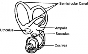
(a) Draw a neat and well labelled diagram of the membranous labyrinth found in the inner ear.
(b) Based on the diagram drawn above in (a), give a suitable term for each of the following descriptions:
(i) The structure responsible for hearing.
(ii) The sensory cells that help in hearing.
(iii) The membrane-covered opening that connects the middle ear to inner ear.
(iv) The nerves that carry impulses from the ear to the brain.
(v) The tube which equalises the air pressure on either side of the ear drum.
Ans:
(a) Below labelled diagram shows the membranous labyrinth found in the inner ear:
(b)
(i) Cochlea
(ii) Organ of corti
(iii) Oval window
(iv) Auditory nerve
(v) Eustachian tube
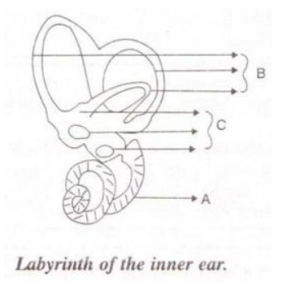
Que-6: Given below is a diagram of a part of the human ear. Study the same and answer the questions that follow:
(i) Give the collective biological term for Malleus, Incus and Stapes.
(ii) Name the parts labeled A, B and C in the diagram.
(iii) State the functions of the parts labeled ‘A’ and ‘B’.
(iv) Name the audio receptor region present in the part labeled ‘A’.
Ans:
(i) Ear ossicles
(ii) A – Cochlea, B – Semicircular canals, C – Ear ossicles
(iii) Cochlea helps in transmitting impulses to the brain via the auditory nerve. Semicircular canals help in maintaining dynamic equilibrium of the body.
(iv) Organ of Corti
Que-7: Draw a labeled diagram of the inner ear. Name the part of the inner ear that is responsible for static balance in human beings.
Ans:
The utriculus (utricle) and sacculus (saccule) are parts of the inner ear responsible for maintaining static balance (posture and position of the head at rest) in humans.
Que-8: Have a look at the posture of this woman who is reading a book and answer the questions which follow:
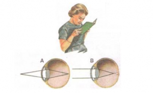
(a) Name the problem she is facing.
(b) What are the two conditions shown in sections A and B of the eye as applicable to her?
(c) What kind of looking glasses she needs?
Ans:
(a) Myopia
(b) A-Normal eye, B-Myopia
(c) Looking glasses with the concave lens are required here.
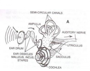
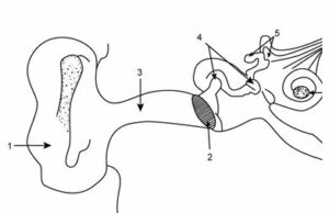
Que-9: The figure given below shows the principal parts of a human ear. Study the diagram and answer the following questions.
(a) Label the parts 1 to 8.
(b) State the role of parts 6, 7 and 8.
(c) Why is it harmful to use a sharp object to remove ear wax? Mention the number and name of the part involved.
Ans:
(a)
1: External ear (pinna),
2: Ear drum (tympanum),
3: Auditory canal,
4: Malleus,
5: Semicircular canals,
6: Cochlea,
7: Auditory nerve,
8: Eustachian tube.
(b)
Part 6 (Cochlea) – It contains sensory cells for hearing.
Part 7 (Auditory nerve) – It transmits impulse of hearing to the brain.
Part 8 (Eustachian tube) – It equalizes air pressure on both the sides of the tympanum.
(c) It is harmful to use a sharp object to remove ear wax as it can rupture the ear drum.
The part involved is part 2 – Ear drum (tympanum).
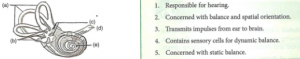
Que-10: Given below is the internal structure of the human ear. Match the structures marked (a) to (e) with their correct function.
Ans:
(a) Semicircular canals: Concerned with balance and spatial orientation.
(b) Ampulla: Contains sensory cells for dynamic balance.
(c) Utriculus and Sacculus: Concerned with static balance.
(d) Auditory nerve: Transmits impulses from ear to brain.
(e) Cochlea: Responsible for hearing
–: End of Sense Organs Class 10 Structured / Applications Concise Biology Selina Ch-11 solutions :–
Return to : Concise Biology for ICSE Class 10 Selina Solutions
Please share with your Friends if helpful
Thanks