Circulatory System Class 10 Concise Structured Applications Type ICSE Biology Selina Solutions Ch-8. In this article you will get the solutions / answer of Structured Applications Skill Type Questions / Problems with figures. Visit official website CISCE for detail information about ICSE Board Class-10 Biology.

Circulatory System Class 10 Concise Structured Applications Type ICSE Biology Selina Solutions Ch-8
| Board | ICSE |
| Subject | Biology |
| Class | 10 |
| Book | Selina Concise |
| Chapter-8 | Circulatory System |
| Topics | Solutions of Structured/Applications/Skill Type Ans Questions |
Structured Applications Skill Type Questions with Answer
Page 111-112
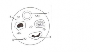
Que-1: Given below is a diagram of a smear of human blood. Study the same and answer the questions that follow:
(a) Name the parts 1, 2, 3 and 4 indicated by guidelines.
(b) Mention two structural differences between the parts labeled 1 and 2.
(c) What is the main function of the parts labeled 1, 2 and 3 respectively?
(d) What is the life span of the part labeled “1”?
(e) Name a soluble protein found in “4” which helps in clotting of blood.
Ans:
(a)
1 → Red Blood Cell (RBC),
2 → White Blood Cell (WBC),
3 → Blood Platelet
4 → Blood Plasma.
(b) The red blood cells are minute biconcave disc-like structures whereas the white blood cells are amoeboid.
(c) Function of part 1 (RBC): Transport of respiratory gases to the tissues and from the tissues, transport of nutrients from the alimentary canal to the tissues.
Function of part 2 (WBC): WBCs play major role in defense mechanism and immunity of the body.
Function of part 3 (Blood Platelet): Blood platelets are the initiator of blood clotting.
(d) The average life span of a red blood cell (RBC) is about 120 days.
(e) Thromboplastin
Que-2: Robin was suffering from blood cancer. Due to his reduced immunity, he has become prone to various kinds of infectious diseases. Answer the questions that follow:
(a) Write the technical term for the above mentioned disease.
(b) Mention the specific structure/cell of the blood whose number increases manifold in this disease.
(c) Write two main functions of the above mentioned structure/cell of the blood.
Ans:
(a) Leukemia
(b) White Blood cells (WBC)
(c) Two functions of WBC
- Helps in fighting with infections.
- Helps in removing damaged tissues and in cleaning up cellular waste.
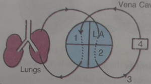
Que-3: Given below is a highly schematic diagram of the human blood circulatory system.
(a) Which part (state the number) represents the heart? Give reason in support of your answer.
(b) Which numbers represent the following respectively?
Aorta
Hepatic portal vein
Pulmonary artery
Superior vena cava
Renal vein
Stomach
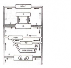
Ans:
(a) The structure 3 represents the heart. It forms the centre of double circulation and is located between the liver and the head (as per the diagram). Also the blood circulation (indicated by 1) begins from heart to lungs.
(b)
| Aorta | 5 |
| Hepatic portal vein | 7 |
| Pulmonary artery | 1 |
| Superior vena cava | 9 |
| Renal vein | 8 |
| Stomach | 10 |
Que-4: The figures given below show diagrammatic cross-sections of three kinds of blood vessels.
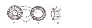
(a) Identify the blood vessels A, B and C.
(b) Name the parts labeled 1-4.
(c) Mention two structural differences between A and B.
(d) Name the kinds of blood that flow through A and through B respectively.
(e) In which one of the above vessels referred to in (a) above does the exchanges of gases actually take place?
Ans:
(a) A- Artery, B-Vein, C-Capillary
(b) 1 – External layer made of connective tissue
2 – Lumen
3 – Middle layer of smooth muscles and elastic fibres
4 – Endothelium
(c) An artery has thick muscular walls and a narrow lumen. It does not have any valve. A vein on the other hand has thin muscular walls and a wider lumen. It has valves to prevent backflow of blood.
(d) A (Artery)- Oxygenated blood, B (Vein)- Deoxygenated blood
(e) At the capillary level the actual exchange of gases takes place.
Que-5: The diagram given below represents the human heart in one phase of its activity. Study the same and then answer the questions that follow:
(a) Name the phase
(b) Which part of the heart is contracting in this phase? Give a reason to support your answer.
(c) Name the parts numbered 1 to 6.
(d) What type of blood flows through the parts marked ‘1’ and ‘2’?
(e) How many valves are closed in this phase?
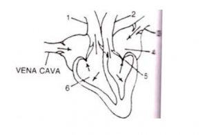
Ans:
(a) Atrial Diastole and Ventricular Systole
(b) Ventricular muscles are contracting during this phase because the valves between the two ventricles and pulmonary artery and aorta are open while the atrio-ventricular valves are closed.
(c)
| 1 | Pulmonary Artery |
| 2 | Aorta |
| 3 | Pulmonary Vein |
| 4 | Left Atrium |
| 5 | Bicuspid Valve |
| 6 | Right Ventricle |
(d) Part 1 (Pulmonary artery) → Deoxygenated blood
Part 2 (Aorta) → Oxygenated Blood
(e) Two i.e., bicuspid and tricuspid valves are closed in this phase.
Que-6: Study the following diagram carefully and then answer the questions that follow:

(a) Name the cell labelled 1.
(b) Identify the phenomenon occurring in A.
(c) Mention two structural differences between 1 and 2.
(d) Name the process occurring in B and C and state the importance of this process in the human body.
Ans:
(a) 1 – Red blood cell
(b) Diapedesis
| RBC | WBC |
| They lack a nucleus. | It have a nucleus. |
| They are biconcave and disc-shaped. | It has a spherical and have different sizes. |
(d) The process which occurs in B and C is phagocytosis. In this process, the WBCs engulf the foreign particles and destroy them, thus preventing the occurrence of disease.
Que-7: Given diagram is a schematic representation of the circulatory system in humans. Study the same and answer the questions that follow :
(a) Label the parts 1 and 4 indicated in the diagram.
(b) Which of the above mentioned number is the thickest artery? Also write its name.
(c) Mention the number and chamber of the heart which has the thickest muscular wall.
(d) Which of the above numbers/structures has the maximum number of blood capillaries?
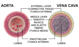
(e) Draw neat and labelled diagrams of the transverse section of vena cava and the part numbered as 3. Make sure to show the structural differences between these two in the diagram.
Ans:
(a) Parts 1 and 4 are:
1. Right Auricle
4. body parts
(b) 3, Aorta
(c) 2, Left ventricle
(d) Lungs
(e) Labelled diagrams of the transverse section of vena cava and aorta showing their structural differences are given below:
Que-8: Given alongside are diagrams of a certain category of blood vessels showing the role of a special structure in their walls. Study the figure and answer the questions that follow.
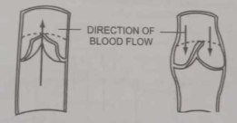
(a) Name the kind of blood vessels shown in the figure. What are its branches termed as ?
(b) Name the structure shown inside the blood vessels. Write its important role.
(c) What kind of blood flows through these blood vessels normally? Name the blood vessel which carries blood from the heart to the lungs.
(d) Name a similar kind of blood vessel which is related to the liver and kidney.
(e) Draw a neat and labelled diagram of the transverse section of the blood vessel shown above showing the three layers of its wall and lumen.
Ans:
(a) The kind of blood vessels shown in the figure is vein. Its branches are termed as venule.
(b) The structure shown inside the blood vessels are valve. Its role is to prevent the backflow of blood.
(c) Deoxygenated blood flows through these blood vessels normally. The blood vessel which carries blood from the heart to the lungs is the pulmonary artery.
(d) Hepatic vein and renal vein are related to Liver and Kidney respectively.
(e) Below diagram shows the transverse section of a vein with the three layers of its wall and lumen labelled:
Que-9: Given below is a diagram of the external features of the human heart. Study the figure and answer the questions that follows :
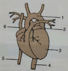
(a) Label the guidelines shown as 1 to 6 in the figure.
(b) Write the important role of parts 5 and 6.
(c) Name the chamber of the heart which collects blood from the lungs through a blood vessel. Also write the name of the blood vessel.
(d) Write one structural and one functional difference between the blood vessels 4 and 5.
(e) What happens when there is a blockage in any coronary artery or any of their branches?
Ans:
(a) Labelled guidelines are:
1. Aortic Arch
2. Left atrium
3 Left ventricle
4. Aorta
5. Inferior vena cava
6. Superior vena cava
(b) Inferior vena cava transports deoxygenated blood from the posterior or the lower region of the body (including abdomen and legs) to heart and superior vena cava transports deoxygenated blood from the anterior or upper regions of the body (including head, chest and arms) to the heart.
(c) Left Atrium collects blood from Lungs. The blood vessel involved is Pulmonary vein.
(d) One structural and one functional difference between Inferior vena cava and Aorta is:
| Inferior vena cava | Aorta |
|---|---|
| Thin wall and wide lumen. | Thick wall and narrow lumen. |
| Transports deoxygenated blood to heart. | Transports oxygenated blood from heart to body parts. |
(e) When there is a blockage in any coronary artery or in any one or more of their branches, there is deadening of the corresponding area of heart muscles leading to myocardial infarction (i.e., heart attack).
–: End of Circulatory System Class 10 Concise Structured Applications Type ICSE Biology Selina Ch-8 solutions :–
Return to : Concise Biology for ICSE Class 10 Selina Solutions
Please share with your Friends if helpful
thanks

7 thoughts on “Circulatory System Class 10 Concise Structured Applications Type ICSE Biology Selina Solutions”
Thank u
keep in touch for any icse /isc related query
Amazing answers
Can u give the answers for all the chapters
https://icsehelp.com/concise-biology-icse-class-10-solutions-selina-publishers/
Amazing answers
Can u give the answers for all the chapters
after lock down over in india genetics answer will be uploaded other chapter are available
yes your all chapter uploaded after lock down over in India